

A. Introduction
- three families: - Lycopodiaceae, Selaginellaceae, Isoetaceae
- vascularized leaves, roots and stems
- exarch protestelic vascular tissue
- xylem - tracheids only
- phloem - sieve cells
- stem has endodermis and pericycle
- sporangia associated with sporophylls
Lycopodium | SelaginellaIsoetes |
Homosporous - One type of spores - Bisexual gametophytes - Gametophyte develops outside the spore wall | Heterosporous - Two types of spores: megaspores and microspores - Megaspores: female; microspores: male - Gametophyte develops inside the spore wall |
No ligules | Has ligules |
B. Lycopodiaceae

Lucopodium "club mosses". They are not the awesome mosses but want to be like them..so they joined the "club".

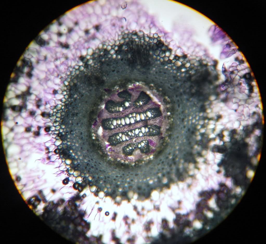
A cross section of the stem of Lycopodium is made.

Cross section of Lycopodium that is stained with toluidine blue.
The stem is an exarch plectostele (a type of protostele).
Another Tol blue staining.

Cross section of Lycopodium that is stained with phloroglucinol.

Strobili of Lycopodium.

Sections through the strobilus of Lycopodium is viewed.

All the spores in the sporangia of Lycopodium is the same size.
Lycopodium is homosporous.
C. Selaginellaceae

Pots of Selaginella krausiana are sacrificed for Science.

The roots of S. krausiana grows from places where branches are.


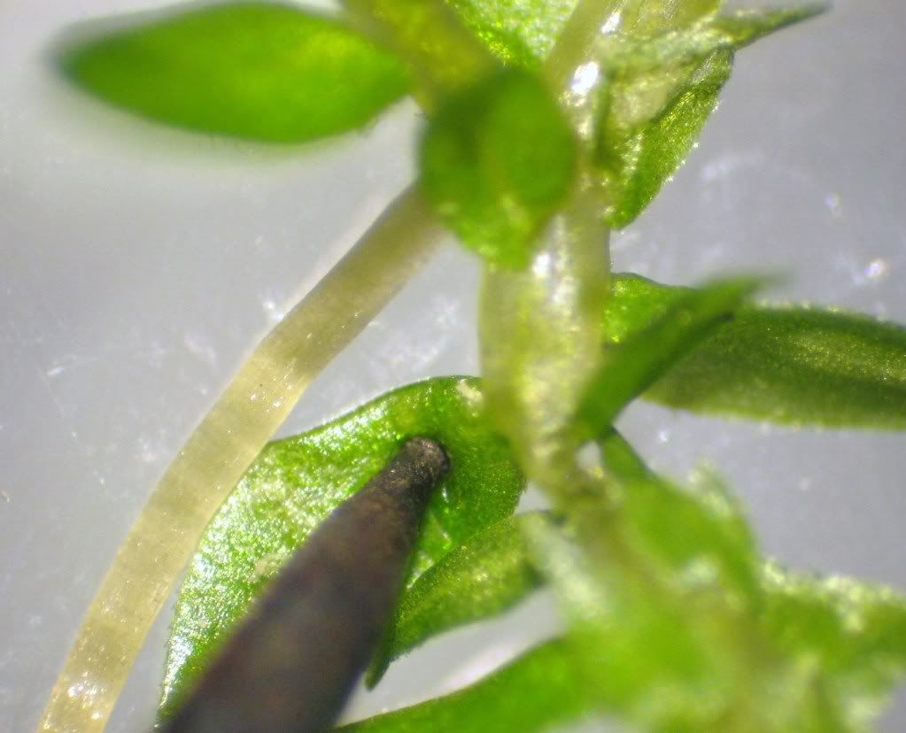
Dissection microscope is used to observe the ligule of S. krausiana.
Pull the adaxial side of the leave downwards and reveal the colourless ligule.
The ligule looks like a hair.
It may be secreting water and mucilage to keep the leaf moist.
The leaves arranged in whirl.
Another ligule (the white speck in the axil of leave). This one was found by Debbie L.
 |
| Vegetative stem of S. wallacei. The leaves are isophyllous (same shape and size).  A cross section of the stem of S. wallacei. It has an exarch protostele. |
 |
Reproductive structure of S. wallacei.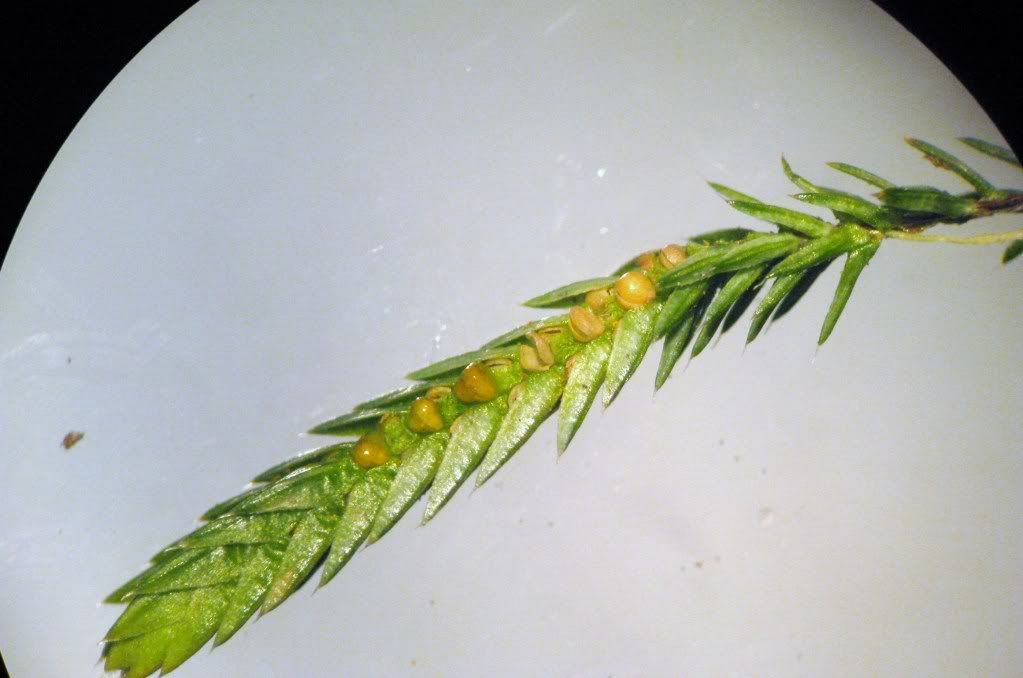 |

Microspore is much smaller than mecrospore.
(I couldn't find my picture of mecrospore. - OYS)

A long-section through the strobilus of of S. wallacei is examined.

There are more microsporangia than megasporangia.
Left- megaspore; right - microspore.

Megasporangium with 4 megaspores (3 of them are showing and 1 is hiding behind).
6.2 Monilophytes
Equisetaceae (Horsetails)
A. Introduction
- One genus, Equisetum, survived.
- Reduced leaves in whorls at the nodes.
- Hollow stem.
- Xylem contains tracheids (some species have short vessels).
- Have sieve cells in phloem.
- Have endodermis and a pericycle in the stem.
- Silicified epidermal cells.
- Conspicuous strobili.
- Homosporous with small, free-living, photosynthetic gametophytes.
B. Equisetaceae

The stem is photosynthetic.

Rhizomes of the Equisetum. Rhizome fragmentation may occur and the plant reproduce vegetatively.

Stomata on the stem of Equisetum.

Equisetum stem cross section.

Equisetum strobilus.

Strobilus under a dissecting microscope.

Sporangiophore is the sporangia (grey) bearing structure.

Sporangia with four elaters under a compound microscope.

Long section through a strobilus. Spores are strained red.

The Equisetum gemetophytes mounts.
-Yee Sing
No comments:
Post a Comment