

Ground Tissue System
I. Parenchyma
---
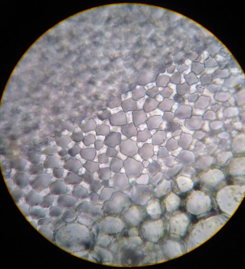
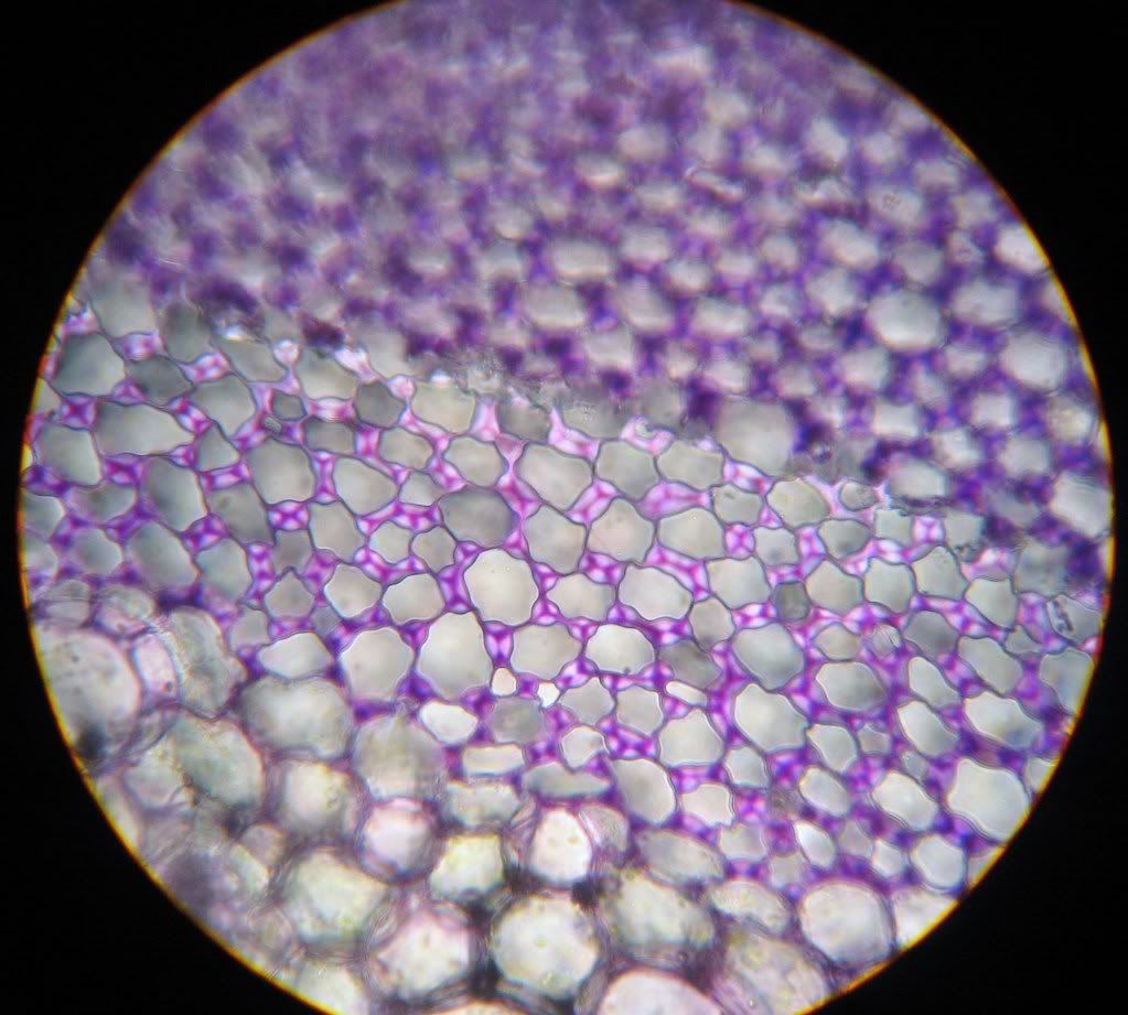
 IKI stained section of an onion (Allium cepa). The large circular cells are parenchyma cells and the red arrow is pointing at a nucleus. One of the many functions of parenchyma cell is storage, but shouldn't starve turn purple when stained with IKI? It turns out the nutrient here is in the form of sugar and not starch.


II. Collenchyma
---

III. Sclerenchyma
---
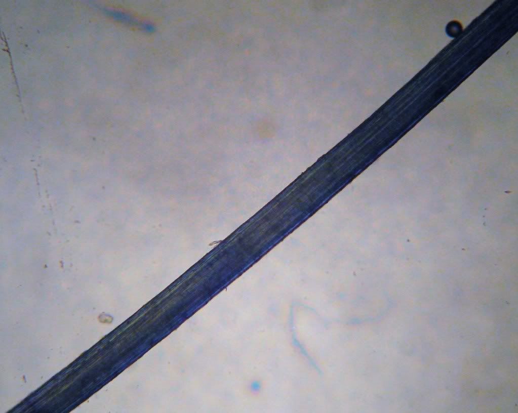
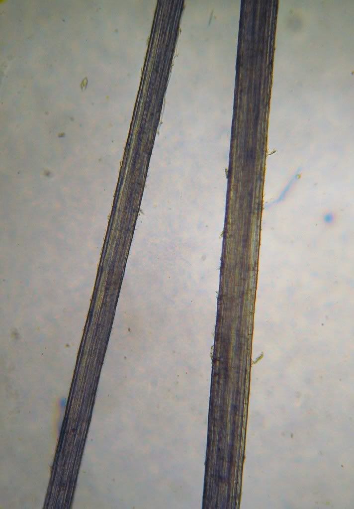 Unstained fibre bundles of Sansevieria (Mother-in-law's tongue).


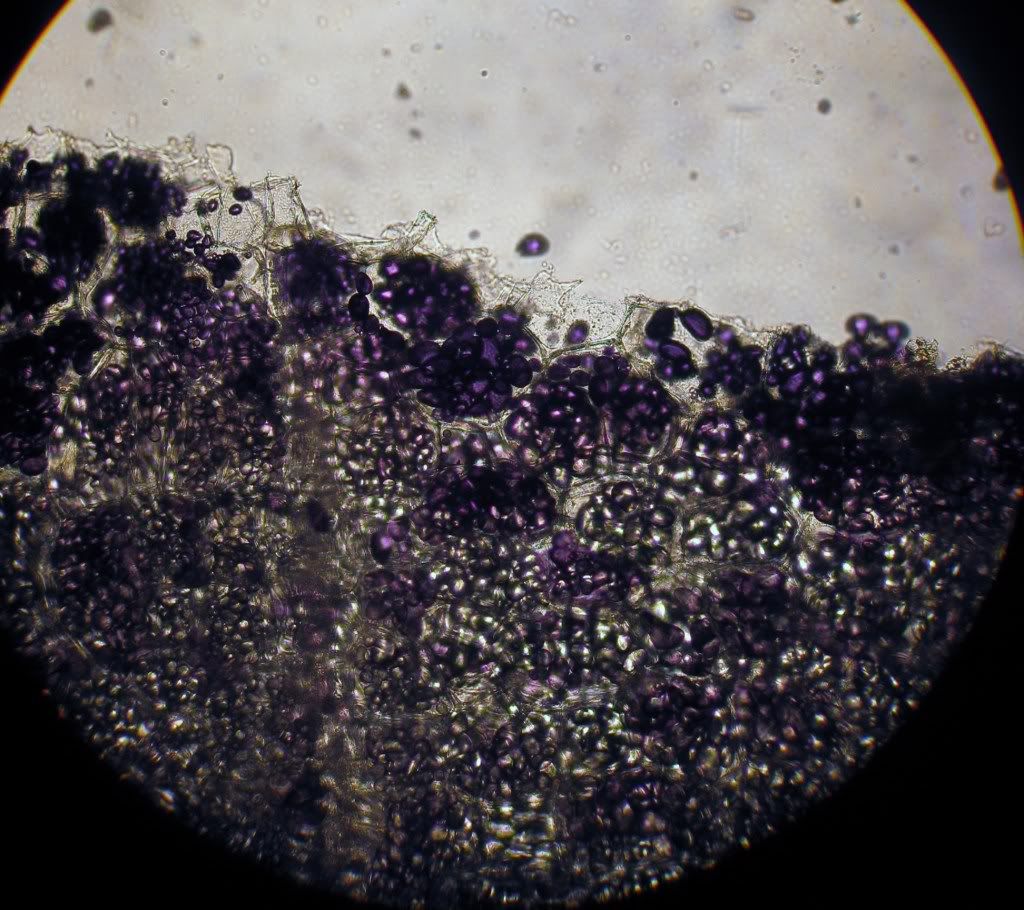
Branched sclereid of Sciadoptiys verticillata (Japanese Umbrella Pine) needle. The sclereids look like little elves hiding behind green bushes.
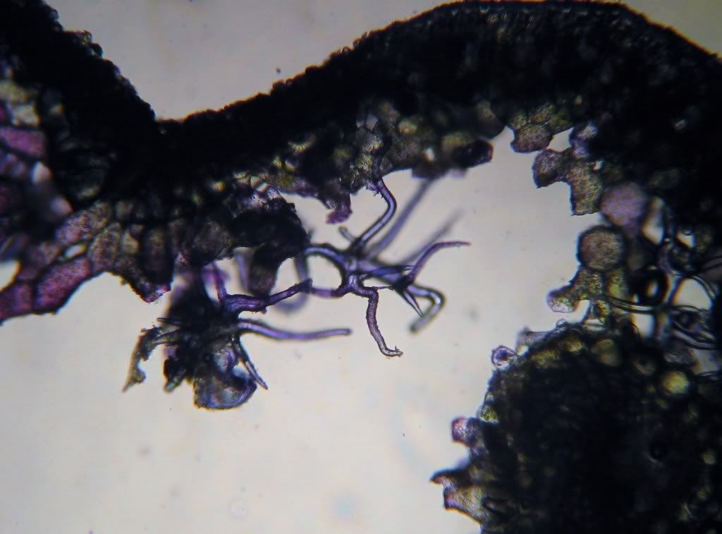 The branched sclereids turn blue after staining with Tol blue indicating their walls are lignified.
Vascular Tissue System
IV. Xylem
---
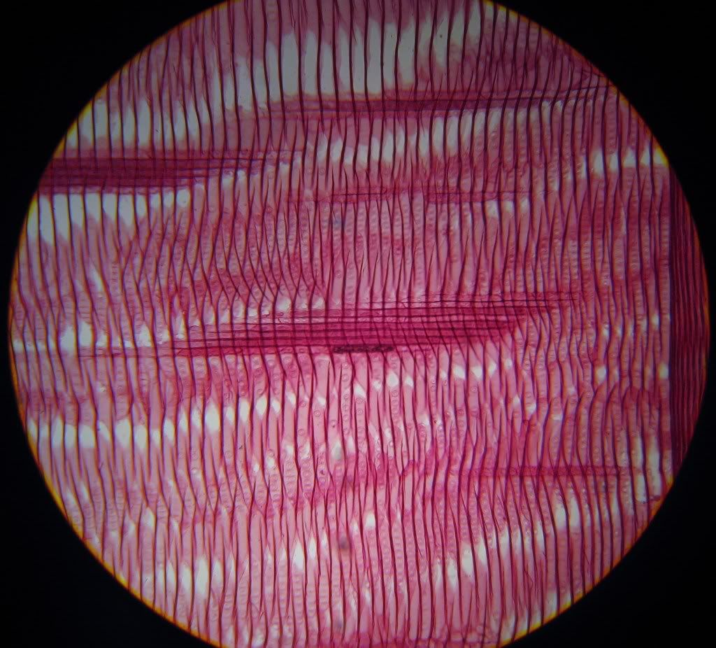 Can you name all the structures in this Pinus (pine) wood section?
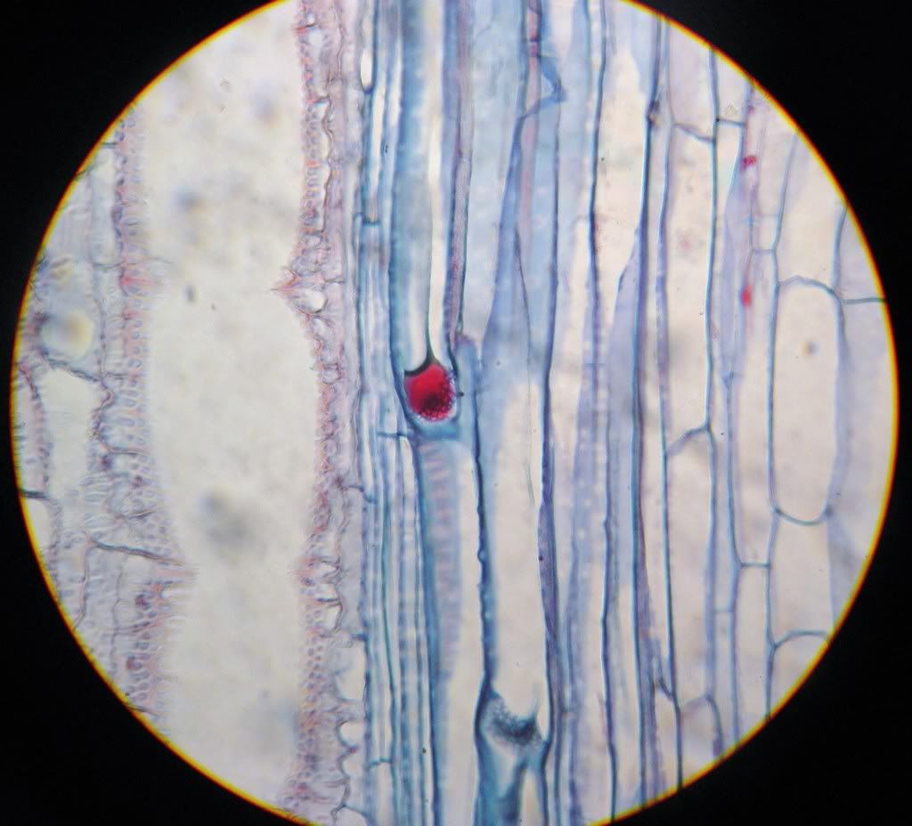
The longitudinally elongating structures are the parenchyma of rays. The long vertical cells with the pores are tracheids and the pores are called 'pits'
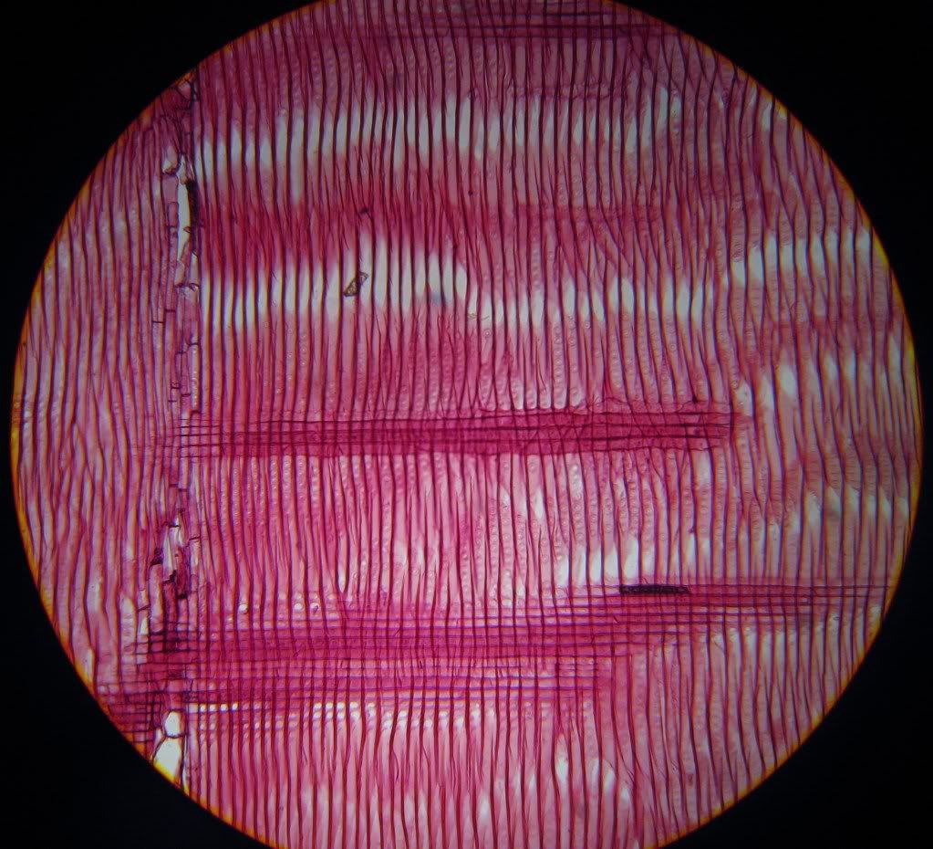 Another longitudinal section of the pine wood.
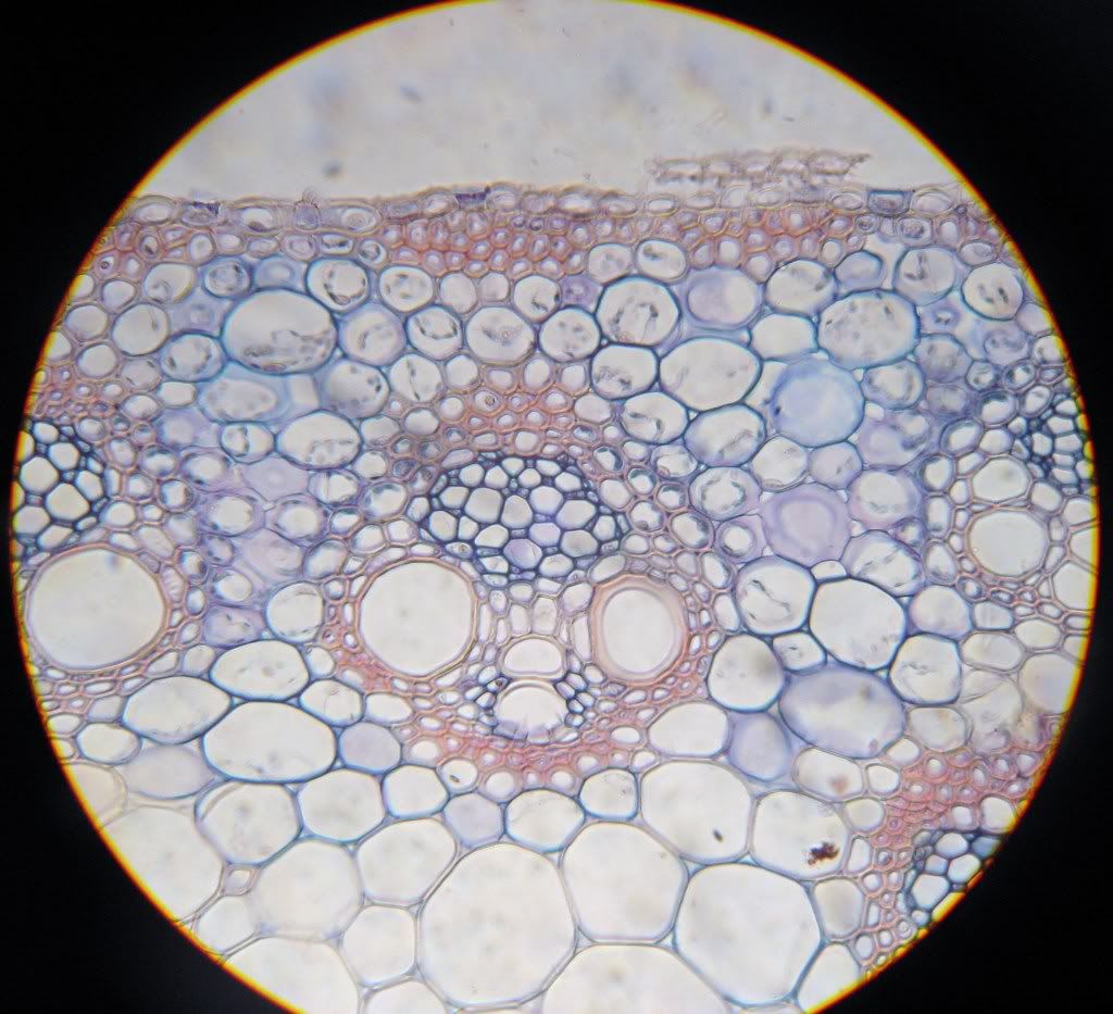
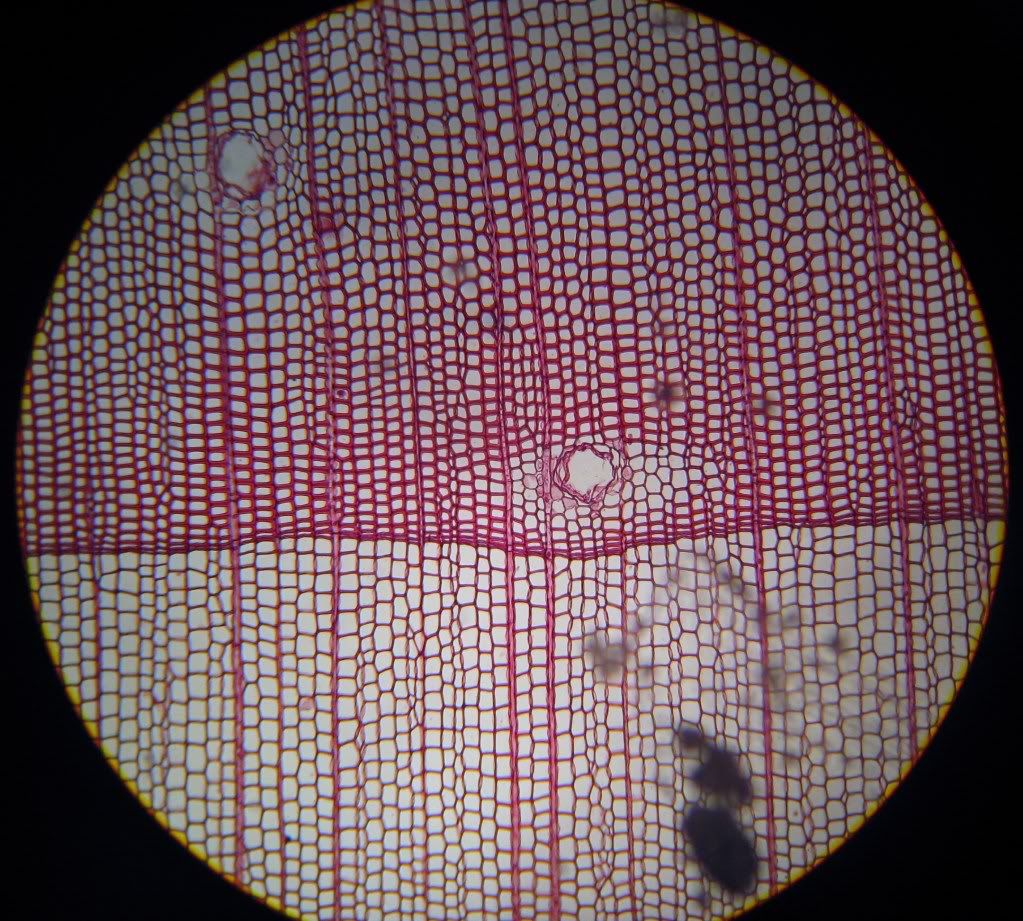 A cross section of the pine wood. Where are the rays in this section? And what are the two big holes (one in the centre and the other on the top left)?
The rays are the thick vertical lines and the holes are actually resin ducts (not covered yet).
The longitudinally elongating structures are the parenchyma of rays. The long vertical cells with the pores are tracheids and the pores are called 'pits'
The rays are the thick vertical lines and the holes are actually resin ducts (not covered yet).
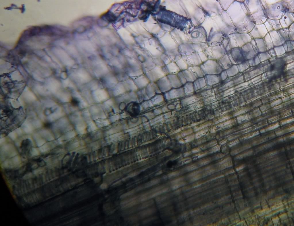
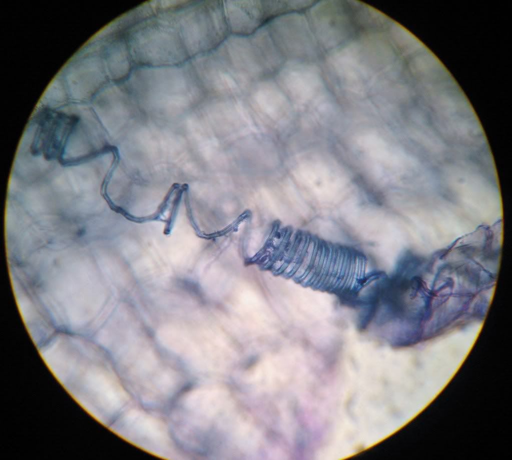
Stained longitudinal sections of Solenostemon scutellarioides (coleus) stem. Check out the helical/spiral secondary walls! The 2 above sections are courtesy of Debbie.
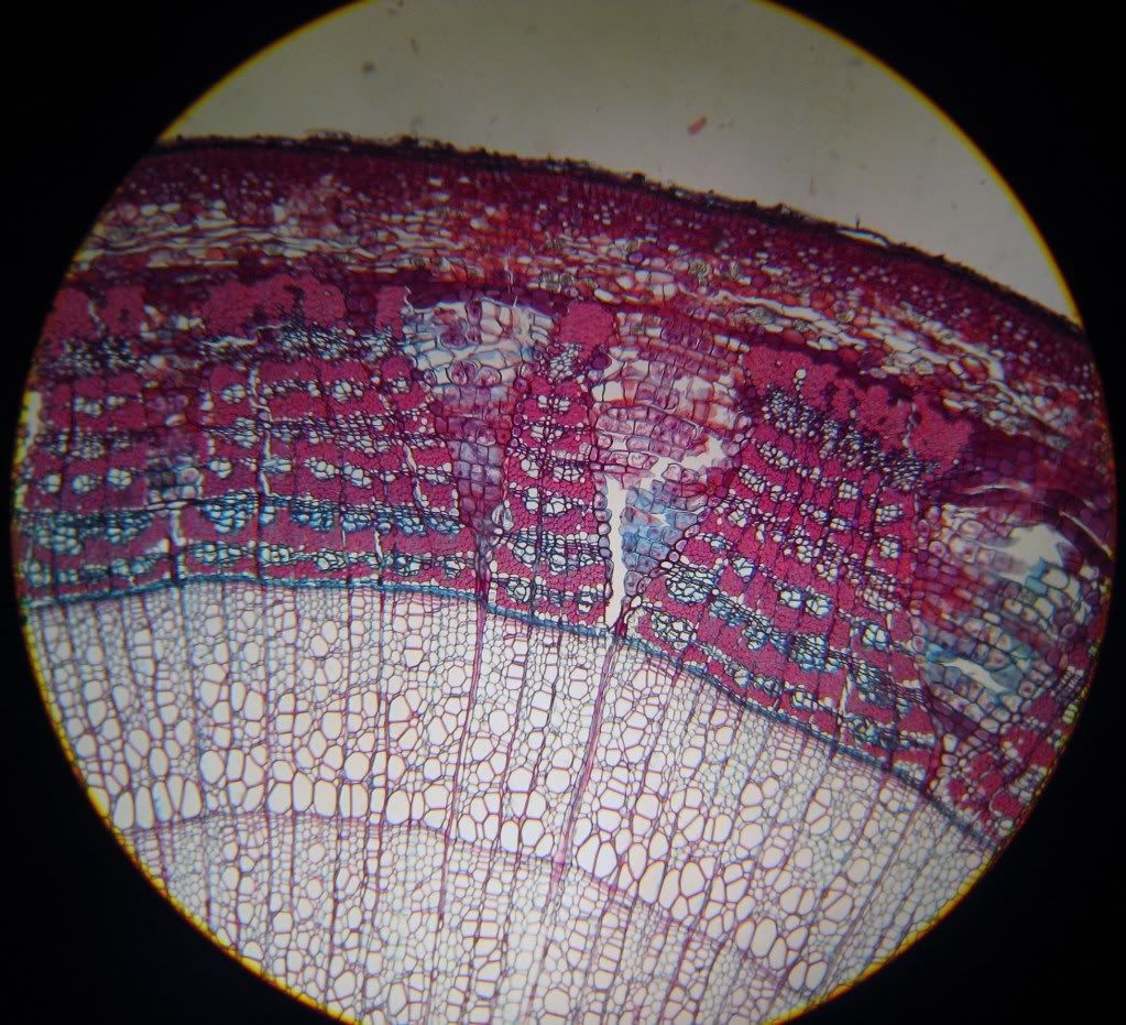
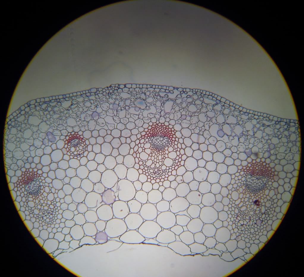 Ranunculus (buttercup) prepared slide section.
V. Phloem
---
 Sieve tube members, companion cells - Zea mays (corn)
Dermal Tissue System
VI. Epidermis
---
---
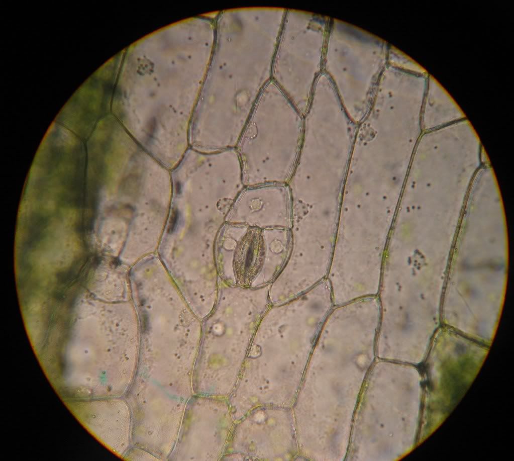
The structure in the centre is the stomatal apparatus and the others are parenchyma cells.
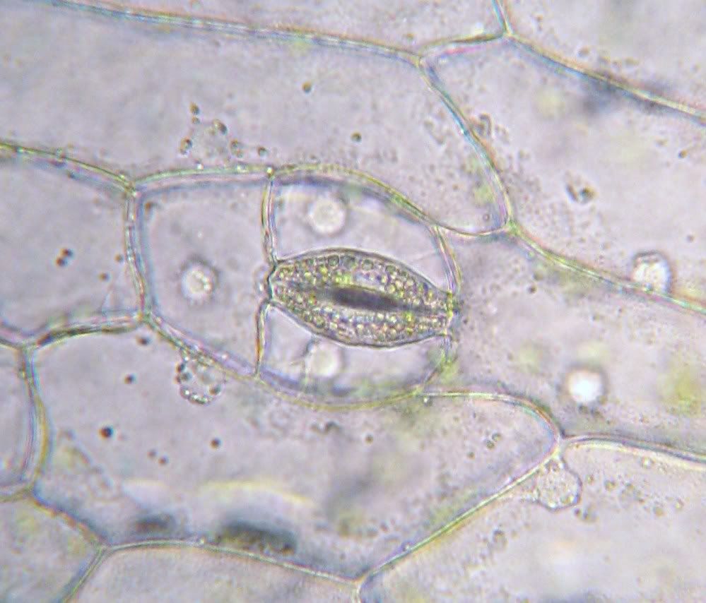
Photo from epidermal peel of Pelargonium (geranium) leaf.
-MIDY
Great pictures, thanks for posting.
ReplyDeleteCould you fix the pictures?
ReplyDelete